Meet the Man “Missing 90% of His Brain”
© Feuillet et al / The Lancet
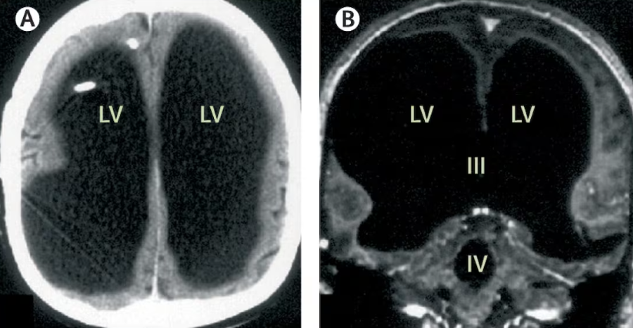
In 2007, a medical case report astonished the scientific community: a 44-year-old man was found to have extremely enlarged brain ventricles and a very thin cortical layer—so much so that media headlines claimed he was “missing 90% of his brain.”
Yet this man reportedly lived a largely normal life: he held a job, had a family, and had only mild neurological complaints. Scientists continue to debate how much brain tissue he truly lost, and how the mind compensates in extreme cases.
How the Discovery Was Made
The man first sought medical help after experiencing mild weakness in his left leg for over two weeks. Imaging scans revealed massively dilated brain ventricles (the fluid-filled spaces in the brain) and almost no visible normal brain structures in many regions.
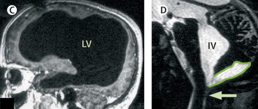
What had been assumed to be brain tissue was largely replaced or compressed by fluid. This was linked to hydrocephalus, a condition where cerebrospinal fluid accumulates and increases pressure within the skull.
He also had a history of treatment for hydrocephalus earlier in life. But over time, the excess fluid appears to have expanded into spaces usually occupied by brain tissue, effectively thinning most of the cortex to a minimal layer.
How “Normal” Was His Life Without a Brain?
Despite what the images showed, the man’s daily functioning was reported as largely intact:
- He worked as a civil servant and supported a family.
- He scored an IQ around 75 on testing—not average, but not severely disabled.
- Socially and behaviorally, he was considered “socially apt” and able to manage everyday tasks.
These findings underscore the brain’s resilience and suggest that in certain conditions, surviving tissue can reorganize functionally.
What We Don’t Know (And Can’t Know Easily)
- It’s uncertain exactly how much neuronal tissue he lost versus how much was merely compressed or displaced. Some scientists argue the “missing” 90% figure is misleading — much of the tissue could have been squeezed into a thinner shell rather than wholly gone.
- We can’t map back all functions easily. Which brain areas were truly destroyed? Which survived and adapted?
- This is a case study, not a controlled experiment. That means we can’t generalize: such adaptation might be extremely rare and dependent on timing, individual biology, and developmental plasticity.
Why This Case Matters
- It challenges assumptions: in neuroscience, the idea that you need “most of your brain” to live is questioned by cases like this.
- It highlights neuroplasticity — the brain’s ability to reorganize and adapt in response to injury.
- It prompts new thinking about consciousness: how much physical tissue does it really require? Some theories suggest awareness is not localized to a single rigid area, but is emergent from networked activity.
- It encourages humility in medical claims: brain imaging is powerful, but interpreting it must be done with caution.
You might also want to read: New Memoir Revives Allegations Against Prince Andrew